How to Read an ECG Strip in 5 Steps
Every rhythm strip, from normal sinus rhythm to complete heart block, can be read with the same short routine. Check the patient, take a quick look at the QRS width, then answer five questions in the same order every time. This page walks through each one, gives the normal values, and points you to free strips to practice on.
Before you start: the patient, then the paper
The golden rule comes first: check the patient, not just the monitor. Monitors name rhythms on their own, and that name is not always right. Loose electrodes and patient movement cause artifact, and a monitor can read artifact as ventricular beats or other abnormal rhythms when nothing is wrong with the heart. A strip only means something next to the person it came from. Watch for signs that the rhythm is affecting them, such as dizziness, confusion, or passing out.
Then know your paper. The standard ECG recording speed is 25 millimeters per second. At that speed the grid becomes a clock:
- One small box (1 mm) is 0.04 seconds.
- One large box (5 small boxes) is 0.20 seconds.
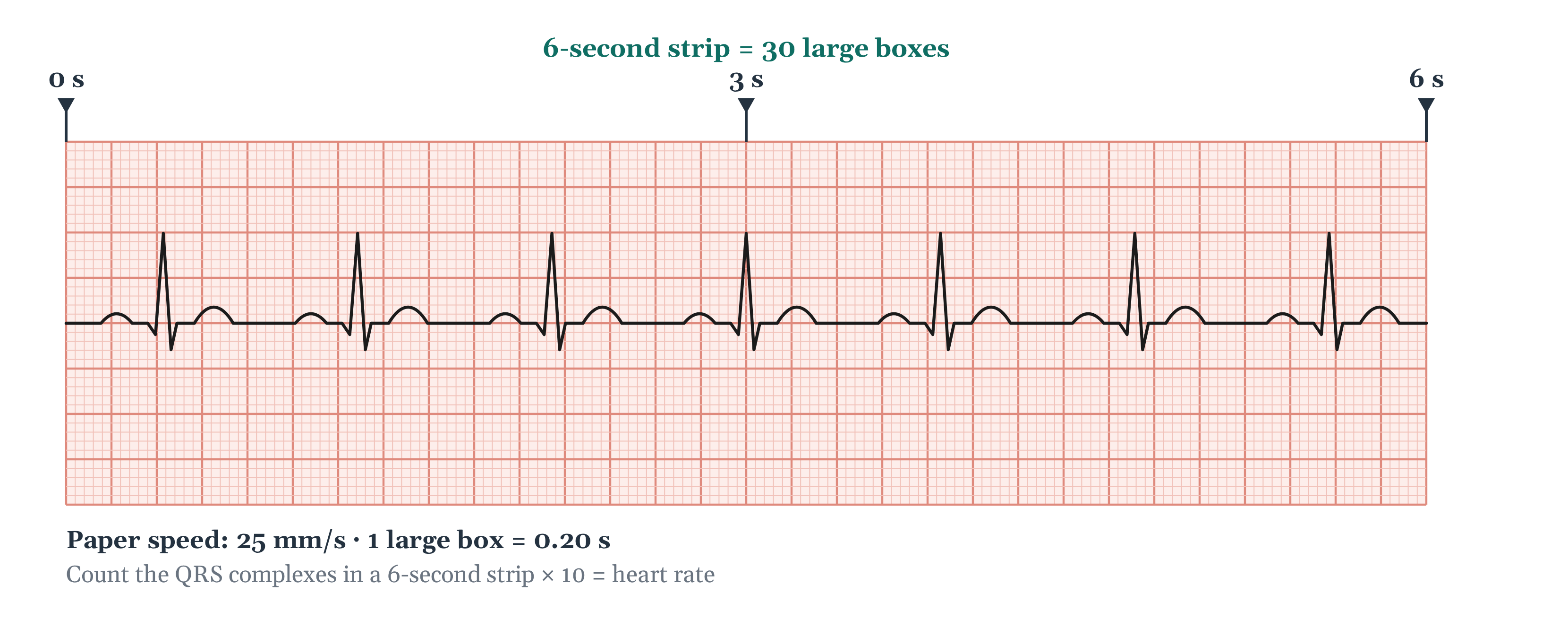
- Five large boxes make one second, so 30 large boxes make a 6-second strip.
Pre-step: is the QRS narrow or wide?
Before the five steps, take one look at the QRS complexes. Are they narrow and sharp, or wide? This is not a measurement yet. It is a first sort that tells you where to pay attention.
A narrow QRS means the impulse traveled the normal path through the ventricles, so the beat started above them. A wide QRS means the beat started in the ventricles themselves, or it started above them and was slowed on the way down, as in a supraventricular rhythm with BBB/aberrancy. You will measure the QRS properly in step 5: 0.06 to 0.12 seconds is narrow, and more than 0.12 seconds is wide.
Narrow or wide is a quick sort, not a measurement. A wide, fast rhythm needs attention right away, and you want to know that before you start counting boxes. The five steps then tell you what the rhythm actually is.
Step 1: Regularity
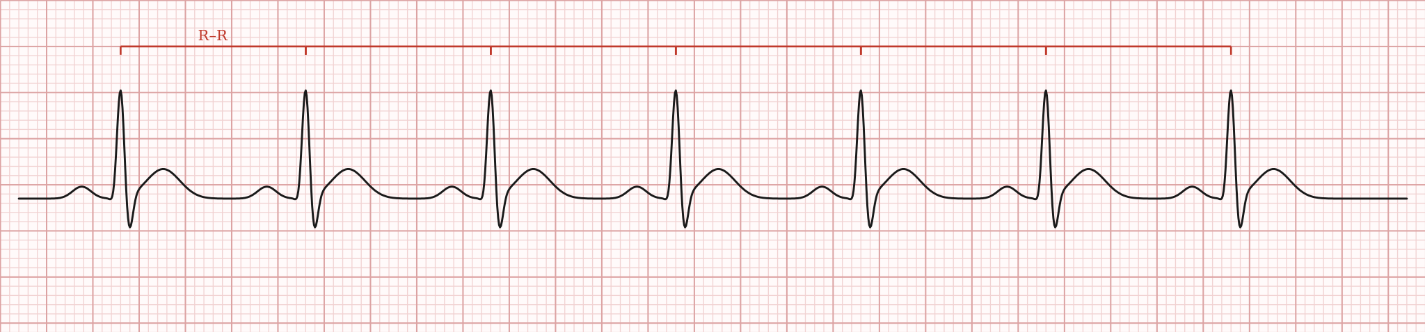
Question: Are the R-R intervals equal?
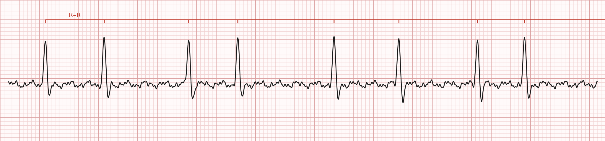
Measure from one R wave to the next with calipers or the edge of a piece of paper, and check across the whole strip, not just two beats. Rhythms fall into four patterns:
- Regular. Every R-R interval is the same. Normal sinus rhythm is regular.
- Irregularly irregular. No two R-R intervals match, and there is no pattern. Atrial fibrillation is the classic example.
- Regularly irregular. The intervals are unequal, but the unevenness repeats in a pattern, as some second-degree heart blocks do.
- Occasionally irregular. The intervals are equal except for an occasional beat that comes early, such as a PVC.
Regularity comes first because it decides how you count the rate in step 2.
Step 2: Rate
Question: How many beats per minute?
For adults, 60 to 100 beats per minute is the normal range taught in rhythm courses and used on this site. Below 60 is bradycardia; above 100 is tachycardia. There are three common ways to get the number:
| Method | How | Use it when |
|---|---|---|
| 6‑second method | Count the QRS complexes in a 6-second strip (30 large boxes) and multiply by 10. | Any rhythm. It is the one to use when the rhythm is irregular. |
| 300 method | Count the large boxes between two R waves and divide 300 by that number. | Regular rhythms only. Quick, but rough. |
| 1500 method | Count the small boxes between two R waves and divide 1,500 by that number. | Regular rhythms only. The most precise of the three. |
Where do 300 and 1,500 come from? At 25 mm/s, one minute of paper holds 300 large boxes, or 1,500 small boxes. Both shortcuts assume every beat is the same distance apart, which is why they give a wrong answer on an irregular rhythm.
If P waves are present, count them the same way to get the atrial rate. The atrial and ventricular rates should match. When they do not, the P waves and QRS complexes are not moving together, and step 3 is where you find out why.
Step 3: P Wave
Question: Is there a P wave before every QRS, upright in lead II?
The P wave tells you where the beat started. Ask four things:
- Are P waves present? No P waves means the beat did not start in the SA node.
- Do they all look the same? A P wave with a different shape started somewhere else in the atria.
- Do they come at a regular rate? Measure P to P the same way you measured R to R.
- Is there one P wave for every QRS? This tells you whether the signal from the atria is reaching the ventricles every time.
In normal sinus rhythm, there is one uniform P wave before every QRS, and it is upright in lead II.
Step 4: PR Interval
Question: How long from the start of the P wave to the start of the QRS?
The PR interval is the time the impulse takes to get from the atria, through the AV node, to the ventricles. A normal PR interval is 0.12 to 0.20 seconds, or 3 to 5 small boxes.
- Longer than 0.20 seconds, with every P wave followed by a QRS, is first-degree AV block. The signal is delayed, not blocked.
- Shorter than 0.12 seconds can mean the impulse is reaching the ventricles faster than normal, as in Wolff-Parkinson-White syndrome.
Measure more than one beat. A PR that stays the same from beat to beat is one finding. A PR that gets longer before a dropped beat, or that has no fixed relationship to the QRS at all, is how the heart blocks are told apart.
Step 5: QRS Duration
Question: How wide is the QRS complex?
Measure from the start of the QRS to the point where it ends. A normal, narrow QRS is 0.06 to 0.12 seconds, up to 3 small boxes. A QRS of more than 0.12 seconds (more than 3 small boxes) is wide. Beats that start in the ventricles, such as PVCs, are wide, and so are beats slowed by a bundle branch block.
This is where the pre-step gets checked with a real measurement. If your first look said narrow and the calipers say 0.14 seconds, trust the calipers.
Normal values at a glance
| Step | Question | Normal |
|---|---|---|
| 1. Regularity | Are the R-R intervals equal? | Regular, with even spacing |
| 2. Rate | How many beats per minute? | 60-100 bpm |
| 3. P Wave | A P before every QRS, upright in lead II? | Yes: upright, one per QRS |
| 4. PR Interval | How long from P start to QRS start? | 0.12-0.20 s (3-5 small boxes) |
| 5. QRS Duration | How wide is the QRS complex? | 0.06-0.12 s (up to 3 small boxes) |
Worked example: the 6-second strip
Go back to the 6-second strip near the top of the page and run the routine.
- Pre-step: the QRS complexes are narrow and sharp.
- Regularity: the R waves are evenly spaced across the strip. Regular.
- Rate: there are 7 QRS complexes between the 0 and 6-second marks. 7 × 10 = 70 bpm.
- P Wave: one upright, rounded P wave before every QRS, and they all look alike.
- PR Interval and QRS Duration: these need calipers on a full-size strip, not a glance at a picture. The measurement drill gives you strips built for exactly this.
Regular, 70 bpm, and a matching P wave before every QRS: this is a sinus rhythm. If the PR and QRS measure normal, it is normal sinus rhythm.
Common mistakes
- Reading the monitor instead of the patient. Artifact can look like a dangerous rhythm. Look at the person first.
- Counting the rate before checking regularity. The 300 and 1500 methods give a wrong number on an irregular rhythm.
- Checking two beats instead of the whole strip. An early beat or a dropped beat can sit anywhere on the strip.
- Eyeballing intervals. A normal PR and first-degree AV block are split by one number, 0.20 seconds. A narrow and a wide QRS are split by another: anything more than 0.12 seconds is wide. Measure.
- Naming the rhythm too early. A strip that "looks like" atrial fibrillation can have P waves you have not found yet. Finish all five steps before you name it.
Reading strips is a skill you build by doing it. These are free, no account needed:
- Chapter 2: the five-step check, the full lesson behind this page.
- ECG basics review: the paper, the waves, and the 6-second rate.
- Find the waves: pick out the P, QRS, and T, and read the box sizes.
- Measurement drill: calipers on the PR, the QRS, and the rate.
- Sinus sampler: name and measure sinus rhythms, then check your answers.
- Free 16-question CRAT Sampler: test yourself.
Want to see what each step is measuring? The conduction engine sample shows the impulse moving through the heart in time with the ECG. The Rhythm Library shows every rhythm as a live monitor, and the glossary defines every term on this page.
Preparing for certification? The CRAT exam guide covers the cost, eligibility, and how the exam is weighted.
Frequently asked questions
What are the five steps for reading an ECG strip?
Regularity, rate, P wave, PR interval, and QRS duration, in that order. Before them comes a quick pre-step: is the QRS narrow or wide? And before everything, check the patient.
Is narrow or wide one of the five steps?
No. It is a pre-step, a quick look at the QRS before you start. It tells you early whether the beat most likely started above the ventricles or in them. You measure the QRS properly in step 5.
What is a normal PR interval?
0.12 to 0.20 seconds, which is 3 to 5 small boxes at standard paper speed. A PR longer than 0.20 seconds with every P wave conducted is first-degree AV block.
What is a normal QRS duration?
0.06 to 0.12 seconds, or up to 3 small boxes. A QRS of more than 0.12 seconds is wide, which points to a beat that started in the ventricles or to a conduction problem such as bundle branch block.
How do you find the heart rate when the rhythm is irregular?
Use the 6-second method: count the QRS complexes in a 6-second strip (30 large boxes) and multiply by 10. The 300 and 1500 methods only work when the rhythm is regular.
Is an ECG the same as an EKG?
Yes. Both are short for electrocardiogram, and the American Heart Association uses both. A rhythm strip is a tracing of at least 6 seconds from one or two leads.
Can I trust the rhythm name on the monitor?
Not on its own. Loose electrodes and patient movement cause artifact, and a monitor can read artifact as ventricular beats or other abnormal rhythms. Look at the patient and read the strip yourself.
The five-step order is the method taught throughout Cardiac Cadence. The paper standards, normal values, and definitions on this page come from these sources:
- Kligfield P, et al. Recommendations for the Standardization and Interpretation of the Electrocardiogram, Part I. AHA/ACCF/HRS, Circulation 2007 (25 mm/s paper speed).
- Open RN, Nursing Advanced Skills, Chapter 7: Interpret Basic ECG, CC BY 4.0, adapted in part from StatPearls (box values, the 6-second method, the four regularity patterns, P wave questions, PR and QRS ranges, artifact).
- StatPearls, EKG Rhythm, NCBI Bookshelf (sinus rhythm P wave and rate).
- Kusumoto FM, et al. 2018 ACC/AHA/HRS Guideline on the Evaluation and Management of Patients With Bradycardia and Cardiac Conduction Delay (first-degree AV block, PR over 200 ms).
- American Heart Association, Electrocardiogram (EKG or ECG).
The strips on this page are Cardiac Cadence's own. This page is for education and exam preparation. Nothing on it is medical advice.